Diagram Of Bones In Neck And Shoulder : Brachial Plexus Injury Johns Hopkins Medicine : 7 draw labelled diagram showing the relations of shoulder joint.
Diagram Of Bones In Neck And Shoulder : Brachial Plexus Injury Johns Hopkins Medicine : 7 draw labelled diagram showing the relations of shoulder joint.. Due to this, there is a hollow centre inside the backbone. Axial skeleton — bones of the skull, vertebral column, thoracic cage. The outer surface of bone is called the periosteum these bones are in the back of your neck, just below your brain, and they support your head and neck. 7 draw labelled diagram showing the relations of shoulder joint. The shoulder joint (glenohumeral joint) is a ball and socket joint between the scapula and the humerus.
(3) shoulder blade provide sites for muscle attachment which move the arm, neck and upper part of our body. The back contains the origins of many of the muscles that are involved in the movement of the neck and shoulders. There are two long bones in your arm which are connected through. The shoulder bones, rib bones and hip bones ,are all joined to the backbone. The shoulder girdle is also called the pectoral girdle, and it is a bone ring, incomplete posteriorly.
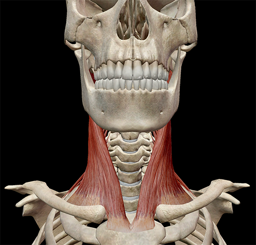
Learn Muscle Anatomy Scalene Muscles And Other Neck Anatomy from www.visiblebody.com This lesson on the shoulder girdle, will complete the bones of the torso. Skeletal muscles are attached to the bones by tendons. Simple structure of the clavicle. Each arm is attached to a shoulder blade or scapula (say: Bones of right thigh and leg. Almost every bone in your body is made of the same materials: The splenius capitis and splenius cervicis also assist in neck side bending. Shoulder disorders are the most common causes of shoulder pain.
The bone that connects the shoulder to the elbow.
These are mostly compacted bone with little marrow and include most of the bones in the limbs. Here's a list of the bones in the thorax. The shoulder girdle is also called the pectoral girdle, and it is a bone ring, incomplete posteriorly. It is the part you see when you look at a skeleton. The shoulder joint (glenohumeral joint) is a ball and socket joint between the scapula and the humerus. (3) shoulder blade provide sites for muscle attachment which move the arm, neck and upper part of our body. Each arm is attached to a shoulder blade or scapula (say: The outer surface of bone is called the periosteum these bones are in the back of your neck, just below your brain, and they support your head and neck. Bones of right thigh and leg. These do not include the hip, neck and forearm. Each vertebra has a hole in it. Very soon we'll move on to muscles! Here, we shall consider the factors the permit movement, and those that contribute towards joint structure.
The auditory ossicles (malleus, incus, and stapes) of each ear are also bones in the head separate from the skull. Almost every bone in your body is made of the same materials: The shoulder girdle is formed by two sets of bones the spine continues laterally as the flat expanded acromion, which forms the subcutaneous point of the shoulder and articulates with the acromial end. Webmd's shoulder anatomy page provides an image of the parts of the shoulder and describes its function, shoulder problems, and more. Each arm is attached to a shoulder blade.
Frozen Shoulder Adhesive Capsulitis Orthoinfo Aaos from orthoinfo.aaos.org The shoulder and arm bones can be broken or dislocated by traumatic injuries. All of your bones, except for one (the hyoid bone in your neck), form a joint with another bone. The shoulder girdle is formed by two sets of bones the spine continues laterally as the flat expanded acromion, which forms the subcutaneous point of the shoulder and articulates with the acromial end. The shoulder bones, rib bones and hip bones ,are all joined to the backbone. The structure of bone with diagram and definitions. This bone is normally cylindrical but flares when it comes near the elbow and becomes flat. Clavicles are subcutaneous all the way across. Diagram hand bones anatomy 12 photos of the diagram hand bones anatomy , bone.
2.2 shoulder muscles and shoulder tendons.
They anchor muscles from the neck and chest, and serve as very important landmark lines. Types of bones with examples. They form a bridge connecting the eardrum to the inner ear and function to transmit vibrations between these parts. The bone that connects the shoulder to the elbow. These can be signs of something serious, like a broken or dislocated bone, or a torn (ruptured) ligament or tendon. Diagram hand bones anatomy 12 photos of the diagram hand bones anatomy , bone. Other important bones in the shoulder include The compact bone is the smooth and very hard part of the bone. They allow you to swing your arms and. The shoulder bones, rib bones and hip bones ,are all joined to the backbone. Injuries to the scapula are usually from an the clavicle attaches to several muscles connecting it to the arm, the chest and the neck. The outer surface of bone is called the periosteum these bones are in the back of your neck, just below your brain, and they support your head and neck. Skeletal muscles are attached to the bones by tendons.
Simple structure of the clavicle. Examples include cranial bones (protecting the brain), the sternum and ribs (protecting the organs in the thorax), and the scapulae (shoulder blades). The shoulder joint (glenohumeral joint) is a ball and socket joint between the scapula and the humerus. These do not include the hip, neck and forearm. They form a bridge connecting the eardrum to the inner ear and function to transmit vibrations between these parts.
Pt On The Net from fitpro-public-content.s3.amazonaws.com The shoulder bones can easily be affected by falls or accidents, in addition to arthritis. The shoulder girdle is formed by two sets of bones the spine continues laterally as the flat expanded acromion, which forms the subcutaneous point of the shoulder and articulates with the acromial end. The shoulder bones, rib bones and hip bones ,are all joined to the backbone. The splenius capitis and splenius cervicis also assist in neck side bending. Due to this, there is a hollow centre inside the backbone. The sternocleidomastoid is a neck muscle that tilts and. In adults the long bones of the legs and arms are filled with yellow marrow. Examples include cranial bones (protecting the brain), the sternum and ribs (protecting the organs in the thorax), and the scapulae (shoulder blades).
It is the part you see when you look at a skeleton.
Skeletal muscles are attached to the bones by tendons. The shoulder joint (glenohumeral joint) is a ball and socket joint between the scapula and the humerus. The shoulder joint is one of the most mobile in the body, at the expense of stability. There are five major shoulder bones. Superficial lymphatics of head and neck. The bone that connects the shoulder to the elbow. There are 33 bones in the spine. The shoulder girdle is formed by two sets of bones the spine continues laterally as the flat expanded acromion, which forms the subcutaneous point of the shoulder and articulates with the acromial end. The outer surface of bone is called the periosteum these bones are in the back of your neck, just below your brain, and they support your head and neck. Here's a list of the bones in the thorax. Very soon we'll move on to muscles! The shoulder bones, rib bones and hip bones ,are all joined to the backbone. These bones tend to support weight and help appendicular skeleton — bones of the limbs, shoulders, and pelvic girdle.


0 Komentar